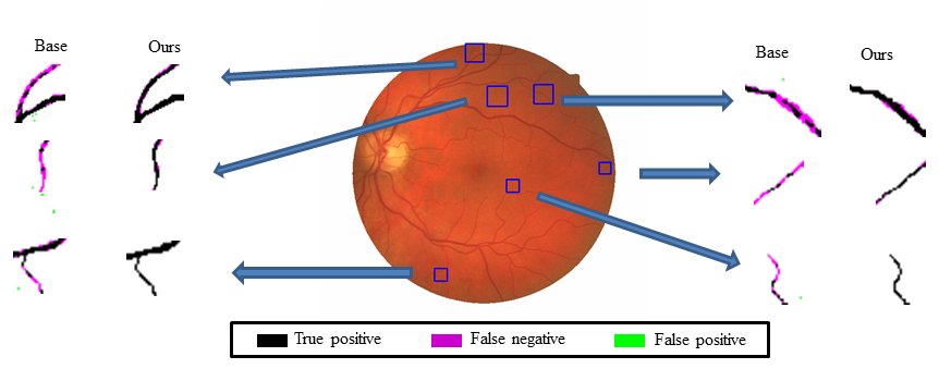
Exemplar segmentation results on 3D neuronal images from the BigNeuron Dataset: Given different base methods such as (left) adaptive enhancement [7] and (right) GWDT [8], our method usually is able to obtain improved results.  Exemplar segmentation results on a retinal image from the DRIVE: compare with the base method such as (left) kernel boost[1], (right) our method produces improved results. The problem of 2D/3D filamentary structure segmentation has a broad range of applications in biological and medical fields. Existing methods often deliver rather erroneous results when encountering small and thin filamentary fragments, where the small fragments often have diverse shapes and appearances, and the backgrounds are usually cluttered and ambiguous. Focusing on this issue, this project aims to boost the performance of the existing base segmenters.
|